
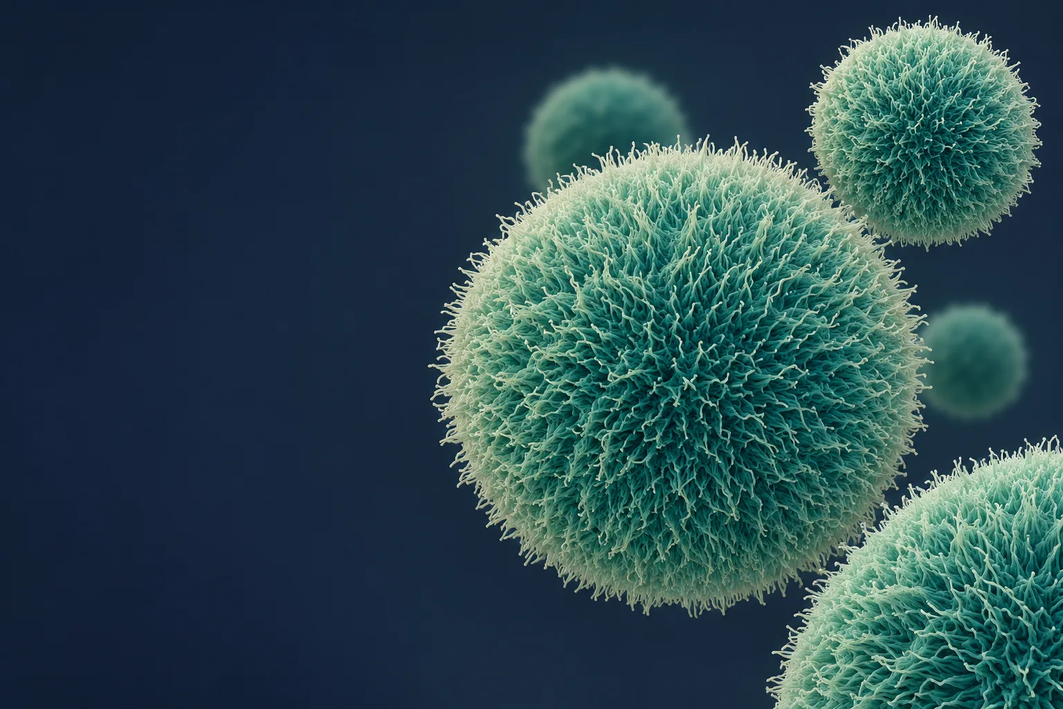
Kuroda, Dezawa and colleagues, in the Proceedings of the National Academy of Sciences.
The Body’s OwnRepair Cells
Muse cells are rare, stress-resistant stem cells found in adult tissue. First described by Professor Mari Dezawa’s team at Tohoku University, they are studied for their ability to sense injury, travel to it and become the cell types needed for repair.
Important
This page is educational and is not medical advice. Muse cell therapies are investigational: none has been approved by the FDA or shown to slow human aging. Vesalius Longevity Labs does not offer Muse cell treatment through this website.
A classic surface marker of human pluripotent cells, used to pick Muse cells out of a mixed population.
Bone marrow, peripheral blood and the connective tissue of many organs.
Transplantation studies have not reported the teratomas seen with some other pluripotent cells.
A Stem Cell Found by Surviving Stress
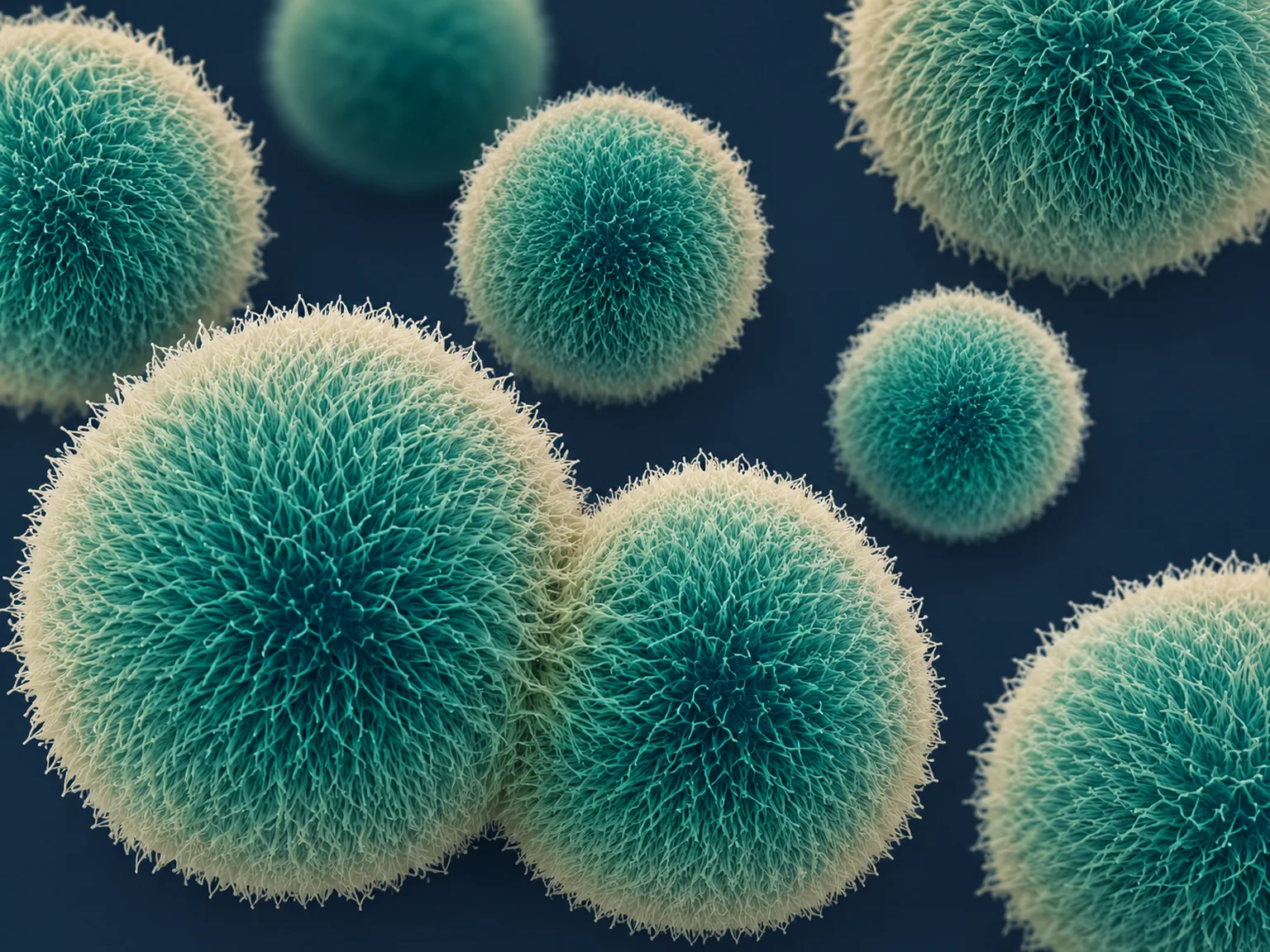
Muse stands for Multilineage-differentiating Stress-Enduring. Dezawa’s team noticed that a small population of human mesenchymal cells survived harsh treatment that killed most of their neighbors. Grown in suspension, those survivors formed clusters that resembled the embryoid bodies made by embryonic stem cells.
The cells express pluripotency genes such as OCT3/4, NANOG and SOX2, and in laboratory studies give rise to cell types from all three germ layers: ectoderm (such as neurons and skin cells), mesoderm (such as heart muscle) and endoderm (such as liver cells).
Unlike embryonic stem cells or iPS cells, Muse cells are not created in a lab. They already exist in the body, need no genetic reprogramming, and show low telomerase activity, which researchers cite as one reason they have not been seen to form tumors.
Sense, Travel, Replace, Integrate
Preclinical studies describe a repair sequence that sets Muse cells apart from most cell therapies, which tend to act mainly through the factors they secrete.
-
01
Damage Sends a Signal
Injured tissue releases sphingosine-1-phosphate (S1P), a lipid that acts as a distress beacon.
-
02
Selective Homing
Muse cells carry the S1P receptor 2, so in animal studies, cells given intravenously migrated preferentially to damaged sites rather than spreading at random.
-
03
Replacing Lost Cells
At the injury, studies from the Dezawa lab report that they take up damaged cells and differentiate into the same cell type, from heart muscle cells to neurons.
-
04
Immune Tolerance
Muse cells express HLA-G, an immunomodulatory molecule, which has allowed donor cells to be studied in trials without HLA matching or long-term immunosuppression.
How Muse Cells Compare with Other Stem Cells
| Property | Muse Cells | Embryonic Stem Cells | iPS Cells | Mesenchymal Stem Cells |
|---|---|---|---|---|
| Source | Naturally present in adult tissue | Early-stage embryos | Reprogrammed adult cells | Adult tissue (mixed population) |
| Genetic manipulation | None | None | Required | None |
| Differentiation range | All three germ layers | Pluripotent | Pluripotent | Mainly mesodermal |
| Teratoma risk | Not observed preclinically | Present | Present | Low |
| Homes to injury when infused | Yes in animal studies, via S1P and S1PR2 | No | No | Limited; many are trapped in the lungs |
| Ethical concerns | Minimal | Significant | Minimal | Minimal |
From the Lab Bench to Human Trials
Clinical trials in Japan have tested CL2020, an intravenous product made from donor Muse cells, in conditions where tissue loss drives disability. Results from a randomized, placebo-controlled trial in subacute ischemic stroke were published in 2024 (Niizuma et al.).
Ischemic Stroke
Randomized, placebo-controlled study.
Acute Myocardial Infarction
Exploratory safety study.
Epidermolysis Bullosa
A rare genetic skin disorder.
Spinal Cord Injury
Early-phase study.
ALS
Early-phase study.
Neonatal Brain Injury
Hypoxic-ischemic encephalopathy.
Muse cell therapies remain investigational and are not approved for clinical use in the United States.
Aging Is Damage That Outpaces Repair
Stem cell exhaustion is one of the recognized hallmarks of aging. Over time, tissues accumulate injury while the body’s ability to replace lost cells declines. A repair cell that already lives in the body and seeks out injury is therefore a compelling subject for longevity science.
A link between Muse cells and human lifespan or healthspan has not been established. These are the open questions worth answering:
A Marker of Repair Reserve?
How do the number and repair capacity of Muse cells change with age, and could they serve as a biomarker of regenerative reserve?
Supporting Natural Release?
Muse cells in the blood rise after acute injuries such as a heart attack. Can that natural mobilization be safely supported?
Age-Related Tissue Loss?
Could Muse cell therapies one day address age-related tissue loss in the heart, brain, skin or other organs?
A Note on Responsible Science
Muse cell research is promising but early. No Muse cell therapy has been approved, and none has been shown to slow human aging. Be cautious of clinics offering unproven stem cell treatments; legitimate access is through registered clinical trials.
Selected References
- Kuroda Y, et al. Unique multipotent cells in adult human mesenchymal cell populations. PNAS, 2010.
- Wakao S, et al. Multilineage-differentiating stress-enduring (Muse) cells are a primary source of induced pluripotent stem cells in human fibroblasts. PNAS, 2011.
- Yamada Y, et al. S1P–S1PR2 axis mediates homing of Muse cells into damaged heart for long-lasting tissue repair and functional recovery after acute myocardial infarction. Circulation Research, 2018.
- Dezawa M. Muse cells provide the pluripotency of mesenchymal stem cells: direct contribution of Muse cells to tissue regeneration. Cell Transplantation, 2016.
- Niizuma K, et al. Randomized placebo-controlled trial of CL2020, an allogenic Muse cell–based product, in subacute ischemic stroke. Journal of Cerebral Blood Flow & Metabolism, 2024.
Follow the Science with Us
Researchers and clinicians interested in Muse cell research and regenerative longevity can reach the Vesalius team through the contact form.